Faster cancer-slide AI cuts computing cost by 50 times, Microsoft Research says
Two new open-weight models shrink the hardware needed to scan thousands of cancer tissue slides, bringing large-scale pathology research within reach of labs that can't afford supercomputer budgets.
Key points - GigaPath-Flash, released in 2025, runs at roughly 50 times less computing cost than the original GigaPath model while retaining 97 percent of its predictive performance. - GigaTIME-Flash processes tissue slides approximately 6 times faster and uses around 8 times less GPU memory than its predecessor. - Both models are released under the Apache 2.0 open licence, meaning any researcher can download and use them for free. - Neither model is cleared for clinical use: they are research tools only, not intended for diagnosing or treating patients. - Model weights are available on Hugging Face, the popular repository where researchers share AI models.
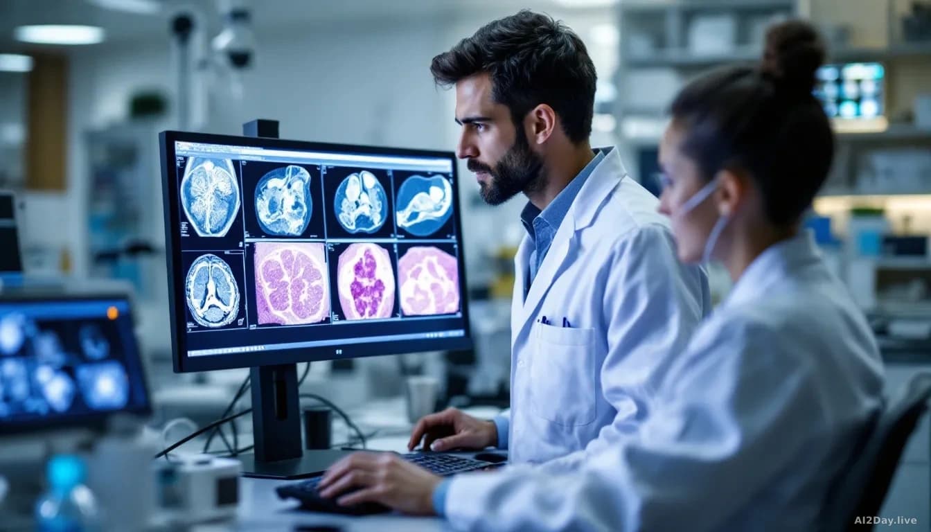
Every time a patient has a cancer biopsy, a pathologist examines a thin tissue slice under a microscope. Hospitals scan those slices into whole-slide images, digital files so detailed they can exceed one billion pixels each. Millions of these images exist, and hidden inside them is information about how tumours behave, which patients respond to which treatments, and why some cancers spread while others don't.
Foundation models, large AI systems pre-trained on enormous amounts of data, have recently shown they can read those signals automatically. The problem has been cost. Analysing a single slide means processing thousands of smaller image tiles, and if a study spans tens of thousands of patients, the computing bill quickly becomes prohibitive.
Microsoft Research now says it has two new models that cut that cost dramatically.
What are these new models, and what do they actually do?
GigaPath-Flash and GigaTIME-Flash are slimmed-down versions of two earlier models the team published in peer-reviewed journals. They do the same jobs at a fraction of the hardware expense.
The original GigaPath, published in Nature in 2024, learned to read entire tissue slides rather than just small patches, capturing both fine cellular detail and the broader layout of a tumour. GigaPath-Flash keeps 97 percent of that performance using a model with 22 million parameters (the internal settings an AI learns during training) instead of roughly one billion. The team achieved this through knowledge distillation, a process where a smaller model is trained to mimic the outputs of a much larger one. We covered a related efficiency technique on 10 August 2026 in "A New Trick Slashes the Cost of Shrinking Giant AI Models".
It takes a routine stained tissue image and predicts what 21 different proteins look like across the tissue, a technique called spatial proteomics mapping. That kind of map normally requires expensive specialised equipment. GigaTIME-Flash swaps out the older image-processing backbone inside GigaTIME for the same compact encoder used in GigaPath-Flash, making the whole pipeline roughly 6 times faster and 8 times lighter on memory, while actually improving accuracy on tissue types the model hadn't seen during training.
How much faster, in practical terms?
The table below shows estimated processing times for generating those protein maps across patient cohorts of different sizes, using a single high-end research GPU.
| Cohort size | GigaTIME-Flash | GigaTIME (original) |
|---|---|---|
| 1,000 slides | ~2 GPU-hours | ~7 GPU-hours |
| 1,000,000 slides | ~70 GPU-days | ~300 GPU-days |
For a single experiment those numbers look incremental. Across a research programme involving repeated hypothesis tests on large datasets, they can be the difference between a study that's feasible and one that isn't.
Should patients or clinicians pay attention?
Not yet, at least not directly. The research team is explicit: these models aren't validated for clinical use, including diagnosis, prognosis or treatment selection. A pathologist should not use GigaPath-Flash to help decide a diagnosis, and no hospital IT team should slot it into a diagnostic workflow.
The longer-term implication for patients is that research studies linking tissue patterns to treatment outcomes could become cheaper and faster to run, which could eventually inform clinical guidelines. Genuinely useful, but years away from the bedside.
What happens next?
Both models are freely available now on Hugging Face. The team describes this as an early research release and notes that broader testing across different scanner types, institutions and patient populations is still needed.
For researchers already working with pathology datasets, the practical step is straightforward: the Apache 2.0 licence lets any academic or commercial lab download the weights and run experiments without restriction.
The thing to watch here isn't whether these models are accurate enough; 97 percent retention is a strong result. It's whether the efficiency gains actually unlock studies that couldn't previously be funded, or whether cost was never the real barrier to population-scale pathology research.
Common questions
What is knowledge distillation?
Knowledge distillation is a training technique where a small model learns to copy the behaviour of a much larger one, ending up nearly as accurate but far cheaper to run.
Do these models replace pathologists?
No. They're research tools designed to help scientists find patterns across large datasets. A trained pathologist still makes all clinical diagnoses.
Can a hospital use these models today?
Not for patient care. The models are released for research purposes only and haven't been tested or approved for clinical decisions of any kind.